
Brain organoids are models derived from pluripotent stem cells to demonstrate brain development in humans. Because they are designed using pluripotent stem cells from patients, all of the genetic information of the patient can be preserved. The organoid model can be used to identify the beginnings of neurodegenerative diseases in patients.
Brain organoids are a more efficient alternative to animal models, which are unable to accurately represent human processes. Brain organoids are also 3D, which means that it is a more representative model of the brain compared to animal and 2D models. Organoids can be developed for several parts of the brain, such as the hippocampus, the hypothalamus, the cerebellum, and more. Though brain organoids include many advantages, the information gathered by them are held back such as hypoxia, stress, and slow maturation.
Real Life Uses of Brain Organoids
A real life example of utilizing brain organoids to identify disease pathways was in 2016, when the Zika virus ravaged Central and South America. Without the use of brain organoids, the connection between the Zika virus and the alteration of neural development in fetuses would not have been found. Additionally, the identification for the development of Alzheimer’s initially used rodent models. However, this was considered lacking in sufficient information.
With brain organoids, research has greatly improved and it is now much easier to locate the start of illnesses like Alzheimer’s. Brain organoids have been found to develop best when surrounded by mouse skin cells (also known as fibroblast feeders); however, this reduces the likelihood of brain organoids mimicking human processes.
In 2024, brain organoids found their way into outer space. Texas Children’s Hospital’s scientists at the Jan and Dan Duncan Neurological Research Institute were sent to the International Space Station. The organoids spent a month in space before landing on Earth. This mission was to evaluate the effect of radiation and gravity on the development of neurological illnesses.
Brain organoids have been used to examine the brain in ways never seen before. Having the ability to detect illnesses before they reach critical levels can save many lives, and constructing 3D models of an accessible brain can allow scientists to evaluate how different environments and harmful substances can alter the development of the brain.
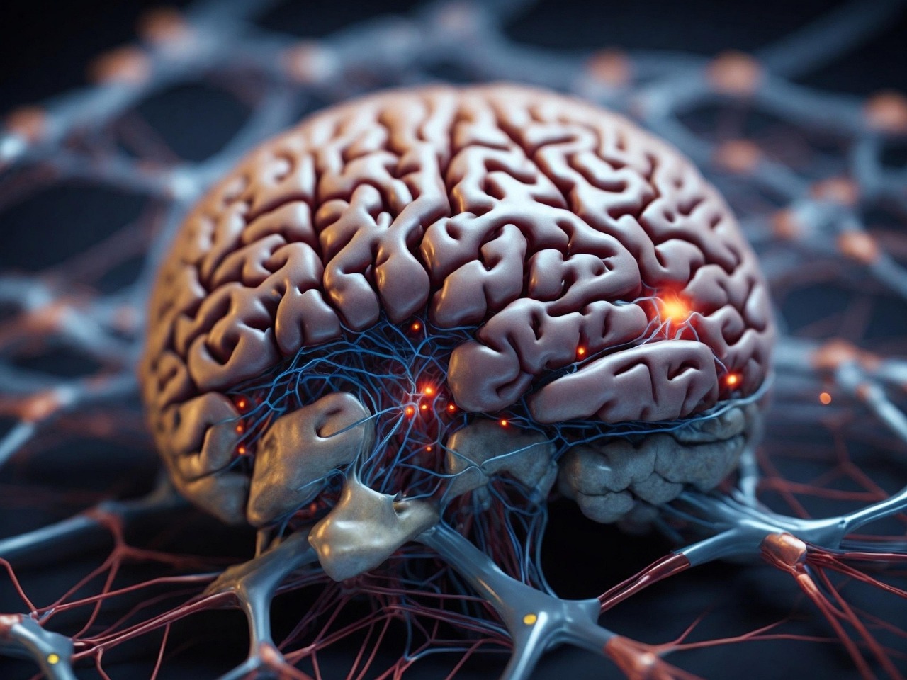
Image Credit: TheDigitalArtist
Sources:
DiCorato, Allessandra. “Brain Organoids Replicate Key Events in Human Brain Development.” Broad Institute, 29 Sept. 2022, www.broadinstitute.org/news/brain-organoids-replicate-key-events-human-brain-development. Accessed 17 Feb. 2025.
Dunlap, Tiare. “Making Lab-Grown Brain Organoids “Brainier.”” UCLA, 29 Sept. 2022, newsroom.ucla.edu/releases/making-mini-brain-organoids-brainier. Accessed 17 Feb. 2025.
Eichmüller, Oliver L., and Juergen A. Knoblich. “Human Cerebral Organoids — a New Tool for Clinical Neurology Research.” Nature Reviews Neurology, vol. 18, no. 11, 1 Nov. 2022, pp. 661–680, www.nature.com/articles/s41582-022-00723-9, https://doi.org/10.1038/s41582-022-00723-9. Accessed 17 Feb. 2025.
Kim, S.Y, and Mi-Yoon Chang. “Application of Human Brain Organoids—Opportunities and Challenges in Modeling Human Brain Development and Neurodevelopmental Diseases.” International Journal of Molecular Sciences, vol. 24, no. 15, 7 Aug. 2023, pp. 12528–12528, www.ncbi.nlm.nih.gov/pmc/articles/PMC10420018/, https://doi.org/10.3390/ijms241512528. Accessed 17 Feb. 2025.
“Texas Children’s Hospital and Baylor College of Medicine Send Human Brain Organoids to the International Space Station | Texas Children’s.” Texas Children’s, 3 May 2024, www.texaschildrens.org/content/news/texas-childrens-hospital-and-baylor-college-medicine-send-human-brain-organoids. Accessed 17 Feb. 2025.
One thought on “Brain Organoids: How Microscopic Models Have Changed the World”